INGROWN HAIR
Ingrown hair (pylonidal sinus) disease
Pilonidal sinus is a cavity (space, cyst) formation in the coccyx area, usually containing a hairball. In fact, the meaning of Pilonidal in the literature is "hair nest". Pilonidal sinus occurs as a result of a chain of events such as pressure, sweating, maceration, inflammation of the hair follicles and ultimately deep penetration, which occur in the coccyx region during hip movements in the presence of a deep natal groove in hairy people. As a result of chronic irritation of the hairs, infection and abscess develop in the space under the skin. A chronic disease occurs as a result of infection or abscess spreading to the skin. Pain, redness and inflammatory discharge on the skin over the coccyx are the most common symptoms. Contrary to popular belief, pilonidal sinus is not a congenital disease but an acquired disease. The disease usually begins from adolescence up to 25 years of age. It is 8-10 times more common in men.
Causes of ingrown hair
* Having too much hair in the area
* Hormones: The secretion of sex hormones produced during the first puberty by affecting the pilosebaceous glands (the onset of pilonidal disease also coincides with this period)
* Narrow and deep groove between the hips (natal cleft)
* Maceration, which occurs as a result of keeping the skin moist, facilitates the ingrowth of the hairs.
* The effect of positive pressure in the natal cavity as a result of obesity or sitting for a long time
* Poor hygiene; not keeping the area clean enough
* Infection: Anaerobic bacteria (especially bacteroids and enterococci) causing inflammation or abscess formation in the hair follicles
Symptoms of ingrown hair
The disease can occur with many different pictures. Symptoms can range from a small sinus opening to a painful mass. Rarely, it may not cause any symptoms in some patients. With the infection, the coccyx area may become red, tender and discharge may occur. The discharge may be clear, dirty yellow, and bloody. If the infection is severe, fever and malaise may occur. Almost all of the patients experience an attack with severe pain, fever, swelling and tenderness, which we can define as acute abscess process. This abscess process often ends with a purulent discharge (pus). This abscess may drain spontaneously. However, surgical drainage is more accurate. A sinus usually develops after the abscess has drained. The sinus is a cavity formed under the skin and opens with one or more openings. A chronic inflammatory discharge may occur from the openings of this sinus. This discharge causes irritation to the surrounding skin over time. Chronic disease causes attacks with pain, swelling and discharge. This event impairs the person's quality of life. The flow rarely stops spontaneously. Surgery is the only option to treat this condition.
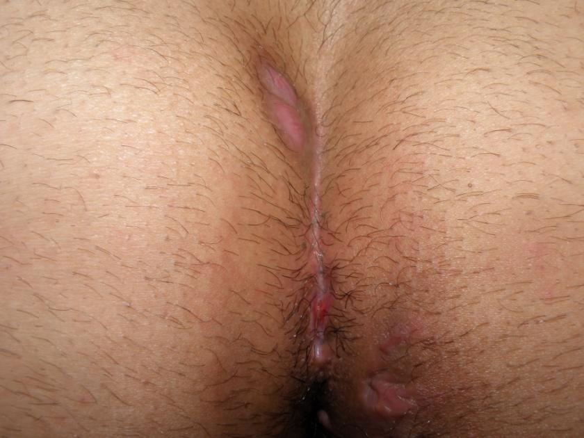
The appearance of ingrown hairs
Treatment of ingrown hair (pilonidal sinus)
Abscess due to ingrown hair
In the presence of an abscess, it should be surgically drained. This procedure can be performed under general anesthesia in the operating room or under local anesthesia in the outpatient clinic. The pus is drained through a small incision. The hairball in the abscess pouch is removed and the debris is cleaned. Clearing the hairball promotes faster recovery and allows for a long period of remission. However, in 60% of patients, the hairs re-accumulate and cause recurrence.
Conservative method
In this method, the opening of the sinus is widened and all the hairs inside are cleaned. A sclerosing agent (80% phenol, silver nitrate or 80% alcohol) is then injected into the sinus. Most commonly 80% phenol is used. Cauterization of the cavity and cryotherapy other than the injection of sclerosing agents can be performed for this purpose. The sclerosing agent injection method is a time-consuming option. Although the average time required for full recovery to occur with the sclerosing effect of the substance is 4-6 weeks, there are cases that are delayed up to 4 months. The recurrence rate has generally been reported below 10% in the literature. However, recurrence rates of up to 40% are also mentioned. It has been reported in some publications that the chance of success is significantly lower in cases with multiple sinus openings and a large cyst or abscess cavity. In some of the cases, the procedure should be repeated at 1-3 week intervals. It can be said that suitable patients for this method are cases with a single sinus opening, without abscess and infection, and with limited volume of tissue. The conservative method is preferred by few clinics. Because the etiological factors causing pilonidal sinus have not disappeared, the recurrence rate is quite high.
Surgical treatment
1. Opening the Pilonidal Sinus and cleaning the hairs (Cystotomy) Only the ceiling of the cavity formed by the hair cysts is opened and the inside is cleaned. The posterior wall (sinus inner wall) of the hair cysts is not removed. It is expected that the cavity formed by the hair cysts, also called the sinus cavity, will be filled spontaneously by dressing frequently. Recovery time is 3-5 weeks on average. If there is a co-morbid infection, antibiotics are administered. The recurrence rate varies between 10-30%.
2. Opening the pilonidal sinus and suturing the sinus edge to the skin (marsupialization) Only the ceiling of the cavity formed by the hair cysts is opened and the inside is cleaned. The posterior wall of the hair cysts is not removed. The wound edges are sutured to the base of the remaining cyst. Thus, the remaining space is reduced. In this method, the patient's daily dressings should be done and the dead tissues and hairs spilled into the wound should be cleaned meticulously. Recovery time is about 4-6 weeks. The recurrence rate varies between 10-25%.
3. Removal of the pilonidal sinus and leaving the wound open The hair cysts are completely removed up to the fascia on the sacral bone with an elliptical incision performed to include the openings of all the hair cysts. The wound is left open. It is expected that the space created by the surgery will be filled by the body by dressing frequently. Recovery time is 4-7 weeks. The recurrence rate varies between 20-25%. The reason for the high recurrence rate is that the factors causing ingrown hair have not disappeared. In addition, it is not preferred today because the healing process is longer and more painful.
4. Removal of the pilonidal sinus and closure of the wound
 After the hair cysts are completely removed, the wound is closed with sutures in such a way that the edges are facing each other. No dressing is required in this method. Recovery time averages 2 weeks. Opening may occur because the wound is partially closed tight. The recurrence rate varies between 6-22%. Because in this method, the factors that cause hair loss are partially eliminated. Shifting the wound sutures to the side instead of the midline (lateral wound closure) halves the recurrence rate.
5. Removal of the pilonidal sinus and closure of the wound with a flap
 It is a very effective method to close the wound with a flap formed from the surrounding tissues after the hair cysts are completely removed. These are called flap surgeries. The flap method has many advantages. Hair cysts and their openings, inflamed skin can be removed widely and the remaining space can be closed without tension with intact tissues. Therefore, recovery is faster. Since the wound is closed and not tight, postoperative pain is less. In addition, it can be prevented that the suture line is in the midline of the body (wounds in the midline of the body heal late). In addition, the narrow and deep slit between the hips, which facilitates the occurrence of the disease, is eliminated with the shifted flap. Thus, the factors causing the formation of pilonidal sinus are eliminated. There are different flap methods. The type of flap to be applied is decided according to the location of the hair cysts, whether they are complicated or not, and the shape of the cavity that will be left behind. The most commonly used is the Limberg flap. The recurrence rate in the Limberg flap technique varies between 1-4%. The Limberg flap technique was modified by us. After this modification, the recurrence rate in our patients decreased to 1%.
Recurrence after surgery
The most important problem encountered after pilonidal sinus treatment is recurrence of the sinus. Recurrence usually occurs within the first 6 months. Recurrence occurs as a result of the healing tissue (scar tissue) being in the midline, infection, insufficient cleaning of the hairs, and re-entering or ingrowing of the hairs. As a result, in addition to the meticulous work of the doctor performing the surgery in the treatment of ingrown hair, the patient must also comply with hygienic conditions. Regardless of which surgery is performed, this area should be regularly cleaned from hair, and body hair should be removed by having a shower every day.
Post-surgical care
If the wound is closed (primary or flap surgeries), the skin should be kept dry and clean until completely healed. If the wound is left open, dressing is necessary to remove the secretions, drainage and discharge in the wound and to allow the wound to heal from the bottom up. During this period and after recovery, the skin of the hip should be kept clean and hair-free. This should be achieved by shaving every 2 or 3 weeks until the age of 30-35 or by using depilatory creams. After the age of 30-35, the hair becomes weak and thin and the depth between the hips decreases. Depending on the preferred surgery and the patient's factors after the surgery, the disease may recur at varying rates. To prevent this, care should be taken to clean the hairs. Depilatory creams or shaving are important to clean the hairs.
A
B
Schematic view of the Limberg flap (A,B)
Sources
1. Tekin A, Balkan M. Plonidal sinusun romboid tranpozisyon ( Limberg) flep ile radikal tedavisi (Radical treatment of the pilonidal sinus with a rhomboid transposition (Limberg) flap). Kolon Rektum Hast Derg (Turkish Jorunal of Colorectal Disease) 1992;2:1820.
2. Tekin A. Pilonidal sinus: experience with the Limberg flap. Colorectal Dis 1999;1:29-33.
3. Tekin A, A simple modification with the Limberg flap for chronic pilonidal disease. Surgery. 2005 Nov;138(5):951-3. 

Assoc. Prof. Ahmet Tekin

General Surgeon